
Surgical Retina Care
Retinal Surgery in Singapore
Retinal surgery aims to repair the light-sensing layer at the back of the eye when it detaches, tears, develops a hole, or is distorted by scar tissue. The aim is to prevent permanent retinal damage and visual loss.
Most procedures are performed as day surgery under local anaesthesia with sedation. Prompt assessment matters because some retinal conditions are time-critical.
At Advanced Eye Clinic & Surgery, retinal surgery is performed by fellowship-trained vitreoretinal surgeon Dr Lee Shu Yen, who previously headed the Surgical Retina department at the Singapore National Eye Centre.
- Setting
- Day surgery for most procedures
- Anaesthesia
- Local anaesthesia with sedation
- Duration
- About 30 minutes to 2 hours, depending on the condition
- Urgency
- Some conditions require same-day assessment and urgent repair
- Vision recovery
- Weeks to months, depending on the condition and timing
- MediSave / insurance
- May apply to medically necessary surgery, subject to the procedure code and plan
Sudden new floaters, flashes or a dark curtain in your vision?
These symptoms can signal a retinal tear or retinal detachment. Because these conditions can be time-critical, seek an urgent eye assessment and contact us immediately for an appointment.
On this page
When Is Retinal Surgery Needed?
The retina works like the film of a camera: it is a thin, light-sensitive layer lining the back of the eye that captures what you see and sends signals to the brain. When a structural problem disturbs the retina, glasses and eye drops cannot repair it.
Surgery may become necessary when:
- The retina detaches from the eye wall
- A hole forms at the centre of the retina
- Scar tissue grows across the macula and distorts vision
- Bleeding inside the eye does not clear on its own
- Scar tissue from advanced diabetic eye disease pulls the retina out of position
Not every retinal condition needs surgery
Conditions such as wet age-related macular degeneration and diabetic macular oedema are generally treated medically with intravitreal injections rather than an operation. Medical retina care at AECS is provided by Dr Lee Shu Yen and A/Prof Kelvin Teo.
Retina Conditions We Treat Surgically
Retinal detachment
A retinal break can allow the retina to separate from the wall of the eyeball. This requires urgent assessment because the detached retina loses its normal blood supply and begins to lose function. The sooner it is reattached—ideally before the central retina, or macula, detaches—the better the potential visual outcome.
Read more about retinal detachment and other conditions on our retinal disease page.
Retinal tears and breaks
A tear caught early can often be sealed with an in-clinic laser procedure, reducing the risk that it progresses to retinal detachment. This is why a sudden onset of new floaters or flashes should be checked promptly.
Macular hole
A macular hole is a small, full-thickness defect at the centre of the retina that can cause blurred or distorted central vision. Surgery can close the hole and support visual recovery.
Epiretinal membrane (macular pucker)
A fine sheet of scar tissue can grow across the central retina, wrinkling it and making straight lines appear wavy. Surgery removes the membrane to relieve the distortion and improve or stabilise vision.
Diabetic retinopathy complications
Advanced diabetic eye disease can cause bleeding into the eye or scar tissue that pulls the retina away from the eye wall. Vitreous haemorrhage and tractional retinal detachment may require surgical repair.
Vitreous haemorrhage
Bleeding into the vitreous gel blocks light from reaching the retina. If the blood does not clear on its own, surgery can remove it and allow treatment of the underlying cause.
Types of retinal detachment
- Rhegmatogenous detachment begins with a tear that lets fluid seep under the retina and lift it away. It is the most common type and is often linked to age-related changes in the eye and myopia.
- Tractional detachment occurs when scar tissue contracts and pulls the retina away, most often in advanced diabetic eye disease.
- Exudative detachment involves fluid building up beneath the retina without a tear, due to inflammation, injury or an underlying disease.
The type of detachment determines whether treatment requires retinal surgery, treatment of an underlying condition, or both.
Types of Retina Surgery
Retina surgery is a family of procedures. The appropriate method depends on the problem being treated and the features of the individual eye.
Vitrectomy
The surgeon removes the vitreous gel that fills the eye to access and repair the retina directly. Vitrectomy is used for retinal detachment, macular holes, epiretinal membranes, diabetic complications and vitreous haemorrhage. A gas bubble or silicone oil may be placed in the eye to support the retina while it heals.

Scleral buckle
A soft silicone band is placed around the outside of the eye to support the eye wall against the detached retina. It is not visible from the front and is generally left in place permanently. This procedure may be preferred for younger patients and certain tear positions, and it can be combined with vitrectomy.
Pneumatic retinopexy
A gas bubble is injected into the eye and positioned so it presses the detached retina back into place. Laser or freezing treatment then seals the tear. This method is suitable only for selected detachments, typically involving tears in the upper part of the retina.
Laser photocoagulation
Laser treatment creates tiny scars that act like spot-welds around retinal tears or leaking blood vessels. It is used to protect existing vision and works best when a tear is treated before it progresses to detachment. Laser photocoagulation is also used in diabetic retinopathy.
Cryopexy
Controlled freezing is applied from outside the eye to seal a retinal tear. It is commonly used with a scleral buckle or pneumatic retinopexy when laser access is difficult.
Intravitreal injections
Intravitreal injections are a non-surgical retina treatment. Medication is delivered directly into the eye for conditions such as wet age-related macular degeneration, diabetic macular oedema and retinal vein occlusion. Learn more about intravitreal injections at AECS.
| Condition | Typical procedure |
|---|---|
| Retinal tear without detachment | Laser photocoagulation or cryopexy, often in clinic |
| Retinal detachment | Vitrectomy, scleral buckle or pneumatic retinopexy, alone or combined |
| Macular hole | Vitrectomy, commonly with a gas bubble |
| Epiretinal membrane | Vitrectomy with membrane peeling |
| Diabetic complications | Vitrectomy, often with laser treatment |
| Wet AMD or diabetic macular oedema | Intravitreal injections rather than surgery |
How Your Surgeon Chooses the Right Procedure
The choice depends on the type and position of the tear or detachment, how much of the retina is involved, whether the vitreous gel is pulling on it, whether you still have your natural lens, your age, and whether this is a first repair or a repeat operation.
Procedures are often combined. A vitrectomy may be performed with laser treatment, or a scleral buckle with cryopexy, because the methods complement one another.
A second procedure is sometimes needed. Most detachments are repaired in one operation, but some eyes require further surgery. Your surgeon will explain how this applies to your individual case.
Macular Surgery for Macular Holes and Epiretinal Membranes
The macula is the small central area of the retina responsible for reading, recognising faces and seeing fine detail. Macular conditions are generally not emergencies in the same way as retinal detachment, but they affect the vision used for detailed tasks.
Both macular holes and epiretinal membranes are treated surgically with vitrectomy. The surgeon removes the vitreous gel, then repairs the hole or removes the membrane at the retinal centre.
Because the macula controls fine central vision, surgery is performed under high magnification and visual improvement is usually gradual over several months. Macular hole repair commonly involves face-down positioning while a gas bubble supports the healing retina.
How Retinal Problems Are Diagnosed
Depending on the condition, your assessment may include:
- Dilated retinal examination — eye drops widen the pupil so the ophthalmologist can examine the retina in detail with a bright light and special lens.
- OCT (optical coherence tomography) — detailed cross-sectional imaging of the macula and retinal layers.
- Ultrasound — used when bleeding or a dense cataract blocks the direct view of the retina.
- Angiography — imaging of retinal blood flow, used in diabetic and vascular cases.
Plan your journey home: dilating drops can blur your vision and increase light sensitivity for several hours, so arrange transport rather than driving yourself.
Concerned about sudden retinal symptoms?
New flashes, floaters, a dark curtain or a sudden drop in vision should be assessed urgently.
What to Expect: From Assessment to Recovery
-
Before surgery
Your surgeon will explain what the examination found, how urgent the condition is, and which procedure is recommended. Bring your medication list and any previous eye records.
If sedation is planned, you may be asked to fast for about six hours beforehand. Follow the specific instructions provided by the clinic.
-
Day of surgery
Most retinal operations at AECS are performed as day surgery. Local anaesthesia removes sensation, while sedation keeps you relaxed. You will not see the surgery itself, although you may be aware of light or vague movement.
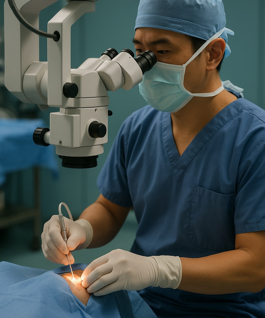
Retinal surgery in progress. The procedure is usually not painful. Depending on the operation and severity of the condition, theatre time can range from about 30 minutes to two hours. Most patients return home after a period of observation in the day ward.
-
After surgery
You will go home with an eye patch or shield and a schedule of eye drops to prevent infection and settle inflammation. Post-operative reviews are commonly arranged for day one, week one and month one, although the exact schedule may vary.
Recovery After Retina Surgery
Recovery varies by procedure, the condition being treated and how quickly treatment began. Your surgeon will give you instructions specific to your eye.
General recovery timeline
First 24–48 hours
Mild grittiness, redness and blurred vision can be expected. Begin your eye drops, rest and avoid strenuous activity.
First week
Vision may remain limited, especially if a gas bubble is present. You may need to maintain a specific head position.
Weeks 2–6
Vision may improve gradually and fluctuate as the eye heals internally. Increase activity only as your surgeon clears it.
Full recovery
Recovery can take weeks to months, and vision may continue improving after the eye has physically healed.
If the macula detached before surgery, or the detachment had been present for some time, vision may not return fully even after the retina is successfully reattached. This is why acting promptly on warning symptoms matters.
Gas bubbles and posturing
If a gas bubble or silicone oil was used to support the retina, vision will be blurred while it remains in the eye. You may be asked to maintain a specific head position, often face-down, so the bubble presses against the repaired area. Follow the exact position and duration given by your surgeon.
Do not fly or travel to high altitude with a gas bubble
Reduced air pressure causes the gas to expand and can raise eye pressure to dangerous levels. Do not fly, travel to high altitude or dive until your surgeon confirms that the bubble has fully dissolved. Tell any doctor, dentist or anaesthetist about the gas bubble before receiving nitrous oxide anaesthesia.
Lifestyle during recovery
- Do not rub the eye, and wear the shield as instructed.
- Use protective eyewear outdoors and avoid dusty environments.
- Avoid strenuous exercise, swimming and heavy lifting until your surgeon clears you.
- Keep diabetes and blood pressure well controlled because they affect retinal healing.
Success Rates and Risks
With current surgical techniques such as vitrectomy or scleral buckle, retinal detachment repair has an anatomical success rate of about 90% after one operation. A second procedure is occasionally needed. Published figures for pneumatic retinopexy generally show a lower single-procedure success rate of around 70–80%.
Successful reattachment and visual recovery are not the same. How much vision returns varies between individuals and depends particularly on whether the macula was involved and how long the retina was detached before repair.
Potential risks
- Retinal re-detachment requiring further surgery
- Cataract progression after vitrectomy, particularly in older patients
- Temporarily raised eye pressure
- Bleeding
- Infection, which is rare
Contact us immediately after surgery if you notice increasing pain, a drop in vision, or a new or expanding shadow.
Cost of Retinal Surgery in Singapore: MediSave and Insurance
MediSave, MediShield Life and Integrated Shield Plans may apply, depending on the surgical code, your insurance plan, panel status, deductibles and co-insurance.
An in-clinic laser or pneumatic procedure generally costs less than a vitrectomy or scleral buckle performed in an operating theatre. Cost also reflects the complexity of the case, whether one or both eyes are involved, and whether a later procedure such as silicone oil removal is planned.
How MediSave and MediShield Life apply
MediSave does not pay a fixed percentage of the bill. Claimable operations are listed in the Ministry of Health's Table of Surgical Procedures, which ranks procedures by complexity and sets withdrawal limits. Retinal procedures span several categories, so the claimable amount varies by case. Day surgery ward charges may be claimable separately, subject to the applicable limit.
The surgical code is assigned based on the procedure performed. In a private setting, MediSave commonly covers only part of the bill, while an Integrated Shield Plan may cover a larger share subject to the policy terms.
What makes up your bill
A retina surgery bill can include the surgeon's fee, anaesthetist's fee, theatre and facility charges, consumables, any implant used, and post-operative reviews. Pre-operative scans, medications and a planned second procedure such as silicone oil removal may be quoted separately.
We will explain which items are included in your quotation and which are billed separately.
Insurance and panel status
Panel arrangements differ between insurers and plans. Our team can verify coverage, advise on pre-authorisation where required, and provide a written estimate before surgery. A detailed cost breakdown will be provided at your consultation before any decision is made.
Frequently Asked Questions
How quickly should I see a specialist for sudden flashes or floaters?
Seek a same-day assessment. Sudden new floaters or flashes can signal a retinal tear. When caught early, a tear can often be treated with laser before it progresses to detachment. A dilated examination is needed to determine the cause.
Is retina surgery painful?
Retina surgery is usually not painful during the procedure because local anaesthesia is used with sedation, although you may notice pressure or movement. Some soreness and grittiness after surgery are normal and can be managed with prescribed drops and simple pain relief.
How urgent is retinal detachment surgery?
Retinal detachment is time-critical. This is particularly important if the macula is still attached because repair before the macula detaches generally offers a better visual outcome. Your surgeon will assess the urgency after examining the eye.
Will my vision fully recover after retina surgery?
Recovery depends mainly on whether the macula was detached and how long the retina had been detached before repair. If the macula was not involved and treatment was prompt, vision may return close to its previous level. If the macula detached or the condition was long-standing, vision may improve without returning fully.
What happens if a retinal detachment is left untreated?
A tear-related retinal detachment typically progresses rather than resolving on its own. As more retina detaches, more vision can be lost, and a longer interruption to the retina's blood supply reduces the potential for recovery. Untreated retinal detachment can lead to permanent vision loss in that eye.
When can I fly after retinal surgery?
If a gas bubble was placed in the eye, do not fly until it has completely cleared and your surgeon has given you permission. Reduced cabin pressure can cause the gas to expand and dangerously raise eye pressure.
How long is recovery?
Day-to-day function often returns over several weeks, while vision can continue improving for a few months. A gas bubble can keep vision blurred and may require specific head positioning. Return-to-work timing depends on the operation and your job.
Is retinal surgery covered by MediSave and insurance?
Retinal surgery is generally medically necessary, so MediSave and insurance may apply. The amount depends on the surgical code, MediSave withdrawal limits, your insurer and plan, deductibles, co-insurance and panel status. Our team can review your specific coverage before surgery.
Will I need cataract surgery after a vitrectomy?
Vitrectomy can accelerate cataract formation or progression, particularly in patients over 50 who still have their natural lens. If cataract surgery becomes necessary, it can be planned with your retinal history in mind. In suitable cases, combined cataract and retinal surgery may be considered.
Can a retinal detachment come back?
Re-detachment is possible even though the first repair succeeds in most cases. Follow-up reviews remain important. If you have had a detachment in one eye, the other eye also carries a higher risk, so any new flashes, floaters or shadows should be treated as urgent.
Our Retina Specialists
Retina care at AECS is provided by two subspecialists covering surgical and medical retina: Dr Lee Shu Yen and A/Prof Kelvin Teo. This allows care to remain coordinated if a condition requires surgery, injections or both over time.

Dr Lee Shu Yen
Senior Consultant Ophthalmologist and Vitreoretinal Surgeon
MBBS, MMed (Ophthalmology), MRCSEd, FRCSEd (Ophthalmology), FAMS
Dr Lee has 24 years of experience and previously served as Head of the Surgical Retina department at the Singapore National Eye Centre. Her clinical interests include retinal detachment surgery, giant retinal tears, retinal complications of high myopia, diabetic vitrectomy, and macular surgery for holes and epiretinal membranes.
She has published 119 scientific papers internationally and served for 17 years as a Clinical Associate Professor at Duke-NUS Graduate Medical School, where she helped establish the ophthalmology programme. She was also core faculty for SNEC's residency and Surgical Retina fellowship training.
Dr Lee was the first woman elected President of the Singapore Society of Ophthalmologists. Her recognitions include the Asia-Pacific Academy of Ophthalmology Distinguished Service Award and a National Day Award from the Ministry of Health.

A/Prof Kelvin Teo
Senior Consultant Ophthalmologist and Medical Retina Specialist
MBBS, FAMS, PhD
A/Prof Kelvin Teo specialises in medical retina, including age-related macular degeneration, diabetic eye disease, retinal vascular disorders and inherited retinal conditions. He spent more than a decade as a senior consultant at the Singapore National Eye Centre and completed medical retina fellowships at SNEC and Sydney Eye Hospital.
He continues active research as an Adjunct Associate Professor at Duke-NUS and a lead researcher at the Singapore Eye Research Institute, with more than 150 peer-reviewed publications and ongoing clinical trial collaborations.
In October 2024, he administered Singapore's first anti-complement therapy for geographic atrophy under early-access approval. Through his research and clinical-trial involvement, suitable patients may have access to emerging retinal therapies where clinically appropriate.
Speak to a retina specialist
Start with a detailed retinal examination at Mount Elizabeth Medical Centre. If your symptoms are sudden, tell our team when you contact us.