
Retinal detachment treatment
Pneumatic Retinopexy in Singapore
Pneumatic retinopexy is a treatment for selected rhegmatogenous retinal detachments. It combines a gas bubble placed inside the eye, laser or freezing treatment around the retinal break, and a prescribed head position while the retina heals.
Whether pneumatic retinopexy is suitable depends on the location and pattern of the retinal break or breaks, the condition of the eye, and whether the patient can follow the positioning instructions.
Not every retinal detachment can be repaired this way, and the decision is made after a dilated retinal examination.
- Type of repair
- Internal gas-bubble repair for selected retinal detachments
- Setting
- Outpatient or day-surgery setting, depending on the clinical plan
- Anaesthesia
- Usually local anaesthesia; additional anaesthesia or sedation may be considered
- Gas type
- SF6, C3F8 or filtered air, chosen by the surgeon
- Positioning
- Individually prescribed head position after the procedure
- Follow-up
- Close early review while the gas remains
On this page
- What is pneumatic retinopexy?
- Who may be suitable?
- How suitability is assessed
- What happens during the procedure
- Gas bubbles and head positioning
- Recovery and follow-up
- Success rates and further treatment
- Risks and warning signs
- Pneumatic retinopexy, vitrectomy and scleral buckle
- Cost, MediSave and insurance
- Frequently asked questions
- Our retina specialists
- References
What is pneumatic retinopexy?
Pneumatic retinopexy is a procedure used to repair selected rhegmatogenous retinal detachments.
It combines three elements: an expanding gas bubble placed inside the eye, sealing of the retinal break with laser photocoagulation or cryotherapy, and a prescribed head position that keeps the bubble supporting the break while healing takes place. [3] [4] [5]
The gas bubble acts as temporary internal support. It floats upward inside the eye and presses against the retinal break, reducing the passage of fluid through the break while the eye absorbs fluid from beneath the retina. Laser or freezing treatment creates an adhesion around the break to support healing.
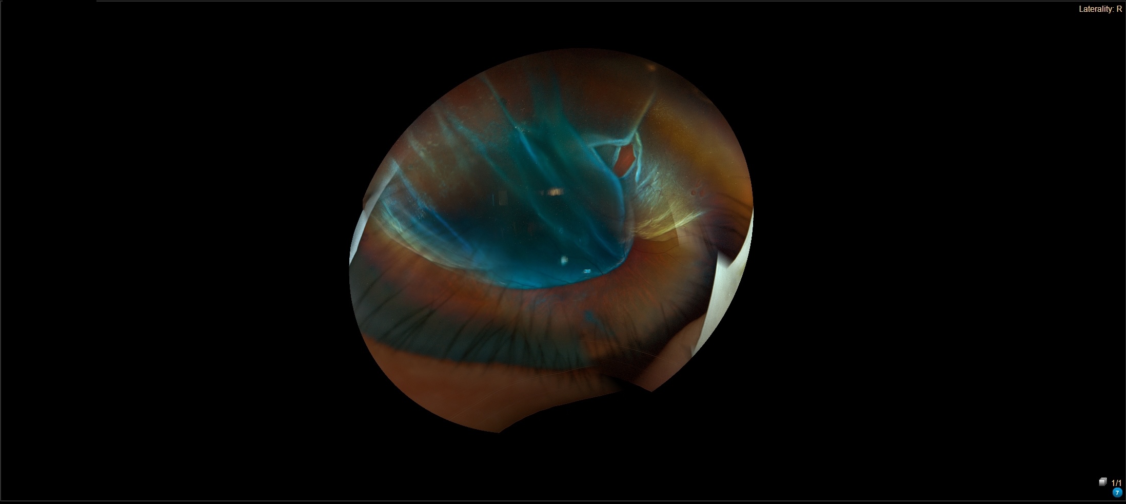
The order of steps is not fixed. Cryotherapy is often applied around the break before the gas is injected. Laser is sometimes applied afterwards, once the retina has flattened enough for the surgeon to treat the break precisely. Both sequences are used in practice, and the surgeon chooses based on the retinal findings.
The gas is absorbed by the body over days to weeks depending on the type used. Removal is not required in normal circumstances.
For the wider picture of retinal detachment repair options, see our retinal surgery page.
Who may be suitable for pneumatic retinopexy?
Suitability is a specialist decision, not something a patient can self-check. The features below are considered during a dilated retinal examination and are weighed together rather than applied as a scorecard. [1] [3] [4]
Features commonly considered favourable
- One retinal break, or a group of small breaks close together, located in the upper part of the detached retina
- A retinal view clear enough for the surgeon to identify every relevant break
- No advanced proliferative vitreoretinopathy or other traction that would resist a gas-only approach
- Ability to hold the prescribed head position and return for close follow-up in the first days after the procedure
- Ability to avoid flying, high-altitude travel and nitrous oxide anaesthesia while the gas remains in the eye
Features that may make another repair more appropriate
- Breaks in positions the gas bubble cannot support reliably, most often in the lower part of the retina
- Widely separated breaks, or breaks that cannot be identified because of vitreous haemorrhage or a dense cataract
- Significant proliferative vitreoretinopathy, marked vitreous haemorrhage, or other complexity
- Eye-pressure or anatomical features that change the risk-benefit assessment
- Inability to posture reliably or attend the required early reviews
- Unavoidable air travel, altitude exposure or planned nitrous oxide anaesthesia during the gas-bubble period
Lens status, the extent of the detachment, macular status and other retinal findings also inform the decision.
Lens status and glaucoma require individual assessment. Aphakia or pseudophakia may affect the likelihood of success, while severe or poorly controlled glaucoma may make pneumatic retinopexy unsuitable. [3] [4]
Evidence context
The PIVOT randomised trial enrolled eyes with a single break, or a group of breaks within one clock hour of detached retina, above the 8 o'clock and 4 o'clock meridians.
These were the trial's recruitment criteria and are not a universal rule for every patient or surgeon. Trial-based criteria help set expectations but do not replace individual clinical assessment. [1]
How suitability is assessed
The assessment focuses on the treatment decision. Broader diagnostic tools are covered on the retinal surgery page.
Your surgeon will typically check the following before recommending pneumatic retinopexy:
- A dilated peripheral retinal examination to locate every break and map the extent of the detachment
- Whether the macula is still attached, since this affects both urgency and expected visual outcome
- Clarity of the cornea, lens and vitreous, and whether all relevant retina can be examined
- Presence of proliferative vitreoretinopathy, vitreous haemorrhage, lattice degeneration or additional breaks
- Eye pressure, lens status, previous eye procedures, and relevant general health
- Whether you can hold the prescribed position, attend close reviews, and avoid flying or altitude while the gas remains
Imaging such as optical coherence tomography or ocular ultrasound is used when it contributes to the individual decision. It is not routinely required in every pneumatic retinopexy case.
Questions worth asking your surgeon
- Why is pneumatic retinopexy being recommended for my eye?
- What are the alternatives, and what would each involve?
- What head position will I need to hold, and for how long?
- Which gas is likely to be used, and how long is it expected to remain?
- What happens if the retina does not remain attached?
What happens during pneumatic retinopexy
Below is a patient-facing sequence. The exact order varies depending on the retinal findings and the surgeon's technique. [3] [4] [5]
-
Final retinal examination
The surgeon confirms the break or breaks, the extent of detachment, and the planned head position. The positioning instructions are explained before discharge.
-
Anaesthesia and preparation
The eye is cleaned and numbed with local anaesthesia. Additional anaesthesia or sedation may be used depending on the patient and the clinical setting.
-
Laser or freezing treatment
Cryotherapy may be applied around the retinal break before the gas is injected. Alternatively, laser may be applied after the gas has been placed and the retina has flattened enough for accurate treatment. Both approaches are used.
-
Gas-bubble injection
A measured amount of expanding gas is injected into the eye to provide temporary internal support. A small amount of fluid may be removed from the front of the eye when the surgeon considers this clinically necessary to manage pressure.
-
Positioning and discharge instructions
Eye pressure and retinal circulation are checked as required. Before discharge, the positioning plan, medication instructions, warning signs and follow-up schedule are explained.
Gas bubbles and head positioning
Why positioning matters
Gas rises inside the eye. The prescribed head angle places the bubble directly against the retinal break, so the required position depends on where the break sits inside the eye.
The surgeon shows each patient the exact position that applies to their eye. Generic clock-face-to-head-angle instructions can mislead because the correct position varies from case to case. [3] [7]
How long positioning is required
Positioning is usually required for much of the day over several days. The exact position, hours per day, allowed breaks, sleeping position and total duration are prescribed individually by the surgeon. Published protocols vary. Follow the instructions given at discharge rather than a general figure found online. [7] [9]
Gas type and how long the bubble lasts
Depending on the case and the surgeon's approach, sulphur hexafluoride (SF6), perfluoropropane (C3F8) or filtered air may be used. The choice depends on the retinal break and the duration of support required. [3] [4]
An exact disappearance date cannot be promised. Your surgeon will tell you which agent was used and when restrictions on flying, altitude and certain forms of anaesthesia can end.
| Agent | Patient-facing description |
|---|---|
| SF6 | A shorter-acting expansile gas. Duration inside the eye varies with concentration, injected volume and the eye itself, usually about 10 to 14 days. |
| C3F8 | A longer-acting expansile gas. It may remain inside the eye for up to 6–8 weeks. |
| Air | Shorter-lasting and non-expansile. Used only when the surgeon judges it suitable for the specific break. |
Recovery and follow-up
Recovery varies from patient to patient. The timeline below is a general guide, not a promise. Your surgeon will give you a plan that fits your eye and your daily circumstances. [5] [7] [9]
The first day
Vision is blurred by the gas bubble. Mild discomfort, grittiness or redness can occur. Begin prescribed drops and follow the head-positioning plan. Arrange for someone to accompany you home if advised.
Early review
Your surgeon checks retinal attachment, gas position and eye pressure. Additional laser may be applied when the retina has flattened, if this is part of your plan.
The first week
Continue the prescribed position and attend every scheduled review. Vision remains affected by the bubble. Review frequency depends on how the retina is settling.
Following weeks
The bubble gradually shrinks. You may notice a moving horizontal line inside your vision as it recedes. Return-to-activity, exercise, work, screen use, washing the eye, sleeping position and driving all follow the surgeon's instructions rather than a fixed calendar.
After the gas has resolved
Vision and any visual distortion may continue to change over weeks to months. Final visual result depends on factors including whether the macula was detached before treatment, how long it was detached, and the overall health of the retina.
Longer-term care
Continue follow-up as advised, and seek urgent assessment if new floaters, flashes or shadows develop in either eye.
Success rates and what happens if further treatment is needed
Published success figures depend on case selection, lens status, detachment complexity, how success is defined, and how long patients are followed. The numbers below come from research studies, not from AECS's own case series, and should be understood in that context.
Findings from the PIVOT randomised trial
The PIVOT trial compared pneumatic retinopexy with pars plana vitrectomy in selected primary rhegmatogenous retinal detachments. The trial reported: [1]
- Primary anatomical success at 12 months of 80.8% after pneumatic retinopexy and 93.2% after pars plana vitrectomy
- Secondary anatomical success after further treatment of 98.7% and 98.6% respectively
- In phakic participants, cataract surgery by 12 months in 16% of the PR group compared with 65% of the PPV group
- Mean visual-acuity and vertical-metamorphopsia results favoured the PR group at specified follow-up points
These figures apply to the trial's eligibility criteria and should not be read as the expected outcome for every retinal detachment.
Findings from a 2023 meta-analysis
A 2023 systematic review and meta-analysis reported higher primary reattachment odds with vitrectomy compared with pneumatic retinopexy across the pooled studies.
Cataract formation or cataract surgery occurred more often after vitrectomy, while new retinal tears occurred more often after pneumatic retinopexy. Each approach carries different risks and may suit different retinal findings. [2]
If the retina does not remain attached
Further retinal detachment repair remains possible if pneumatic retinopexy does not achieve reattachment or a new detachment develops.
Options can include repeat retinopexy, pars plana vitrectomy, scleral buckle, or a combined approach. The next step depends on the reason for failure and the findings on re-examination. [1] [3]
Anatomical success is not the same as visual success. The retina can be reattached without vision returning to its previous level. Final vision is influenced by factors including whether the macula was detached, the duration of macular detachment and the health of the retina.
Risks, complications and when to contact the clinic
Every retinal detachment repair carries risk. Individual risks depend on the eye, the nature of the detachment, general health and the specifics of the procedure. Your surgeon discusses these with you before treatment. The list below is not an exhaustive consent document. [2] [3] [5]
Retinal outcome risks
- Persistent or recurrent retinal detachment
- A missed break, or a new break that develops after the procedure
- Proliferative vitreoretinopathy, which is scarring on the retina that can pull it out of position
- Residual subretinal fluid or retinal folds
Gas-related and pressure risks
- Raised eye pressure
- Gas entering an unintended part of the eye, such as under the retina or into the front chamber
- Multiple small gas bubbles that do not initially combine into one
Other eye risks
- Inflammation, bleeding or infection
- Choroidal detachment
- Cataract progression
- Temporary or lasting visual disturbance
Pneumatic retinopexy, vitrectomy and scleral buckle
The three main procedures for retinal detachment repair work in different ways. The most appropriate option depends on the retinal breaks, the extent of detachment, macular status, lens status, proliferative vitreoretinopathy, other eye findings, and the patient's ability to follow aftercare. [1] [2] [3]
| Consideration | Pneumatic retinopexy | Vitrectomy | Scleral buckle |
|---|---|---|---|
| Basic approach | Gas bubble plus laser or cryotherapy, without removing the vitreous | Removes the vitreous gel and repairs the break, usually with gas, air or silicone oil support | Places an external silicone band or element on the eye wall to support the break |
| Typical role | Selected detachments with break patterns suitable for gas support | Broader range, including detachments that are complex or unsuitable for PR | Selected cases based on age, lens status, break pattern and surgeon judgement |
| Setting and anaesthesia | Often performed in an outpatient setting; anaesthesia is tailored to the patient | Usually an operating-theatre procedure; anaesthesia is tailored to the patient | Usually an operating-theatre procedure; anaesthesia is tailored to the patient |
| Positioning | Central to treatment and usually required | May be required if gas or oil is used | Depends on whether internal gas is also used |
| Lens and refractive considerations | Does not remove the vitreous. Cataract progression was lower than PPV in PIVOT's phakic participants | Cataract progression is an important consideration in phakic eyes | Can change refraction and may cause buckle-related effects |
| Need for further treatment | Lower single-procedure attachment than PPV in PIVOT, with similar final attachment after further treatment | Higher primary attachment than PR in PIVOT's selected population | Evidence and suitability vary; discuss the individual detachment |
See the retinal surgery page for a broader description of each procedure.
Cost, MediSave and insurance in Singapore
Pneumatic retinopexy is listed by Singapore's Ministry of Health under TOSP code SL701R in Table 4A. [10]
MediSave withdrawal limits
MediSave withdrawal limits cap how much can be claimed from a patient's MediSave account for a claimable procedure.
They are not the price of the procedure and not a guaranteed reimbursement amount. Different limits may apply to the surgeon's fee, hospital charges and daily ward charges, and MOH revises these limits from time to time.
Refer to the current MOH published limits for the figures that apply on the day of your procedure, or ask us for a written estimate. [11]
Private-sector surgeon fee benchmark
MOH publishes a private-sector surgeon fee benchmark for SL701R of S$3,000 to S$4,800 before GST (S$3,270 to S$5,232 including GST) for a routine and typical case.
This is a benchmark for the surgeon's fee only. It is not AECS's total package price. Hospital or facility fees, investigations, consumables, medications and follow-up may be billed separately. [10]
MediShield Life and Integrated Shield Plans
MediShield Life and Integrated Shield Plan payments depend on eligibility, the care setting, plan terms, deductibles, co-insurance, pre-authorisation, and the final claim assessment. [12]
Requesting a cost estimate
Ask AECS for an itemised estimate before treatment. Check what is included, what may be billed separately, and how the estimate may change if a further procedure becomes necessary.
See the retinal surgery page for the general MediSave, MediShield Life and Integrated Shield Plan framework.
Not sure which retinal detachment treatment applies to you?
A dilated retinal examination is needed to identify the detachment pattern and the repair options that may be suitable.
Frequently asked questions
What type of retinal detachment can pneumatic retinopexy treat?
Pneumatic retinopexy treats selected rhegmatogenous retinal detachments, meaning detachments caused by a tear or break in the retina that lets fluid pass under it. It is not used for tractional or exudative detachments. Even within rhegmatogenous cases, suitability depends on the position and pattern of the break, media clarity, and the patient's ability to follow positioning instructions.
How does a gas bubble repair a detached retina?
The gas bubble acts as temporary internal support inside the eye. It floats upward and presses against the retinal break, reducing the passage of fluid through the break while the eye absorbs fluid from beneath the retina. Laser or freezing treatment creates an adhesion around the break to support healing.
How will my surgeon decide whether I am suitable?
Your surgeon decides after a dilated retinal examination that maps every break and assesses the detachment. Break position, macular status, media clarity, proliferative vitreoretinopathy, lens status, previous eye surgery, general health, and your ability to hold a prescribed head position and attend close follow-up are all weighed together.
How long must I keep my head in a particular position?
The position and duration are prescribed individually. Positioning is typically required for much of the day over several days after the procedure. Your surgeon will show you the exact position, tell you how many hours a day it applies, and confirm when it can ease. Follow the plan you were given rather than a general figure.
How long does the gas bubble remain in the eye?
The bubble is absorbed by the body over days to weeks. SF6 tends to be shorter-acting; C3F8 lasts longer, sometimes several weeks; filtered air is shorter-lasting still. Exact duration varies with the gas used, the concentration, the injected volume and the eye. Your surgeon tells you which agent was used and when restrictions on flying and certain forms of anaesthesia can end.
What will my vision look like while the bubble is present?
Vision is blurred while the bubble occupies part of the eye. You may see a moving curved or horizontal line at the edge of the bubble, and its position changes as the bubble becomes smaller. This usually resolves as the gas is absorbed.
Can I sleep normally after pneumatic retinopexy?
Sleep positioning is part of your prescribed head-position plan. Some patients are asked to sleep sitting up or on a specific side so the bubble stays over the break. Your surgeon will tell you which sleeping positions are allowed and whether an aid such as a wedge pillow or support cushion is recommended.
When can I fly, drive, exercise and return to work?
Do not fly or travel to high altitude until the gas bubble has fully resolved and your surgeon confirms it is safe. Do not drive until your surgeon advises that it is safe and your vision meets the applicable driving-licence requirements. Return to exercise and work depends on your job and the pace of retinal healing, and follows the instructions given at each review.
What happens if pneumatic retinopexy does not reattach the retina?
Further retinal detachment repair remains possible. Options include repeat retinopexy, pars plana vitrectomy, scleral buckle, or a combined approach. The choice depends on the findings at re-examination. Possible reasons include a missed break, a new break or proliferative vitreoretinopathy.
Can I have nitrous oxide anaesthesia while a gas bubble remains in my eye?
No. Nitrous oxide can expand the intraocular gas bubble rapidly and cause dangerously high eye pressure, with a risk of permanent vision loss. Tell every doctor, dentist, anaesthetist and emergency clinician that you have a gas bubble in the eye. Carry this information with you until the gas has fully resolved. [6]
How does pneumatic retinopexy compare with vitrectomy?
Pneumatic retinopexy uses an intraocular gas bubble without removing the vitreous and is often performed in an outpatient setting. In the PIVOT randomised trial of selected primary rhegmatogenous retinal detachments, pars plana vitrectomy achieved higher primary anatomical success at 12 months (93.2% versus 80.8%). Secondary anatomical success after further treatment was 98.6% and 98.7%, respectively. Among phakic participants, cataract surgery within 12 months occurred in 65% of the vitrectomy group and 16% of the pneumatic-retinopexy group. These trial results apply to the study population and do not determine the most appropriate treatment for an individual eye. [1]
Can MediSave or insurance be used for pneumatic retinopexy in Singapore?
Pneumatic retinopexy is listed under Singapore TOSP code SL701R in Table 4A. Applicable MediSave withdrawal limits and MediShield Life or Integrated Shield Plan benefits depend on eligibility, plan terms, deductibles, co-insurance, pre-authorisation, the care setting and final claim assessment. Ask AECS for an itemised estimate before treatment. [10] [11] [12]
Our retina specialists
At Advanced Eye Clinic & Surgery, retinal conditions are assessed by Dr Lee Shu Yen, a vitreoretinal surgeon and former Head of Surgical Retina at the Singapore National Eye Centre, and A/Prof Kelvin Teo, a medical retina specialist.

Dr Lee Shu Yen
Senior Consultant Ophthalmologist and Vitreoretinal Surgeon
MBBS, MMed (Ophthalmology), MRCSEd, FRCSEd (Ophthalmology), FAMS
Dr Lee is a vitreoretinal surgeon whose clinical focus includes retinal detachment surgery, giant retinal tears, retinal complications of high myopia, diabetic vitrectomy, and macular surgery for holes and epiretinal membranes. She previously served as Head of the Surgical Retina department at the Singapore National Eye Centre.
Her qualifications, clinical interests and professional activities are listed on her profile page.
View Dr Lee's full profile · Book an appointment with Dr Lee

A/Prof Kelvin Teo
Senior Consultant Ophthalmologist and Medical Retina Specialist
MBBS, FAMS, PhD
A/Prof Kelvin Teo specialises in medical retina, including age-related macular degeneration, diabetic eye disease, retinal vascular disorders and inherited retinal conditions. He spent more than a decade as a senior consultant at the Singapore National Eye Centre and completed medical retina fellowships at SNEC and Sydney Eye Hospital.
He continues active research as an Adjunct Associate Professor at Duke-NUS and a lead researcher at the Singapore Eye Research Institute, with more than 150 peer-reviewed publications and ongoing clinical-trial collaborations.
He administered Singapore's first anti-complement therapy for geographic atrophy under early-access approval. His research and clinical-trial work includes emerging retinal therapies.
Need an assessment for a retinal detachment?
A dilated retinal examination is needed to identify the detachment pattern and the repair options that may be suitable. The recommended approach may be pneumatic retinopexy, vitrectomy, scleral buckle or another treatment, based on the clinical findings.
References
The clinical claims on this page are drawn from research studies, professional bodies and official Singapore sources. AECS reviews this page when clinical evidence, professional guidance or regulatory information changes.
- Hillier RJ, Felfeli T, Berger AR, et al. The Pneumatic Retinopexy versus Vitrectomy for the Management of Primary Rhegmatogenous Retinal Detachment Outcomes Randomized Trial (PIVOT). Ophthalmology. 2019;126(4):531–539. doi:10.1016/j.ophtha.2018.11.014
- Roshanshad A, Shirzadi S, Binder S, Arevalo JF. Pneumatic Retinopexy Versus Pars Plana Vitrectomy for the Management of Retinal Detachment: A Systematic Review and Meta-Analysis. Ophthalmology and Therapy. 2023;12(2):705–719. doi:10.1007/s40123-023-00653-9
- Chan CK, Lin SG, Nuthi ASD, Salib DM. Pneumatic retinopexy for the repair of retinal detachments: a comprehensive review (1986–2007). Survey of Ophthalmology. 2008;53(5):443–478. doi:10.1016/j.survophthal.2008.06.008
- American Academy of Ophthalmology. When and How to Use Pneumatic Retinopexy. EyeNet Magazine. June 2019.
- Johns Hopkins Medicine. Pneumatic Retinopexy. Patient information page.
- The Royal College of Ophthalmologists. Ophthalmic Safety Alert: Do not use nitrous oxide when there is gas in an operated eye.
- Moorfields Eye Hospital NHS Foundation Trust. Instructions for post-operative posturing.
- Houston S, Graf J, Sharkey J. Commercial air travel after intraocular gas injection. Aviation, Space, and Environmental Medicine. 2012;83(8):809–810. PMID:22872998
- Hull University Teaching Hospitals NHS Trust. Post-operative posturing following retinal surgery.
- Singapore Ministry of Health. SL701R Retina, Pneumatic Retinopexy. TOSP Table 4A and current private-sector fee benchmark.
- Singapore Ministry of Health. MediSave inpatient and day surgery limits. Current surgical and daily hospital-charge withdrawal limits.
- Central Provident Fund Board. Using MediSave for hospitalisation. General eligibility and claim framework.