
Scleral Buckle Surgery in Singapore
Scleral buckle surgery repairs selected retinal detachments caused by a tear or hole in the retina, known as rhegmatogenous retinal detachment.
The retina is the light-sensitive layer at the back of the eye. A silicone band or sponge is secured to the eye's outer wall to support the retinal break and help the retina reattach.
The buckle is usually used with freezing treatment (cryopexy) or laser to help seal the retinal break. A gas bubble, vitrectomy or both may also be used in selected cases. The recommended approach depends on the detachment and the individual eye.
- Purpose
- Repair selected retinal detachments caused by retinal breaks
- Approach
- Performed from outside the eye
- Setting
- Often day surgery, depending on the case
- Anaesthesia
- Local anaesthesia with sedation, or general anaesthesia
- Buckle
- Usually left in place unless a later problem requires removal
- Recovery
- Physical and visual recovery vary between patients
On this page
What is a scleral buckle?
A scleral buckle is a soft silicone band or sponge secured to the sclera, the white outer wall of the eye. It gently indents the eye wall beneath a retinal tear or hole, bringing the supporting tissue closer to the retina and helping close the break. The buckle sits outside the eyeball, beneath its thin surface covering, the conjunctiva.
The surgeon usually treats the area around the break with freezing or laser. This creates a scar that helps seal the break as the eye heals. Fluid trapped beneath the retina may be drained during surgery or gradually absorbed afterwards. A segmental buckle supports one area; an encircling band goes around the eye. During buckle-only surgery, the vitreous gel inside the eye is usually left in place.[4]
Scleral buckling supports the retina from outside the eye. Vitrectomy removes the vitreous gel from inside the eye to relieve pulling on the retina and allow internal repair. The operations may be used separately or together.
When scleral buckle surgery may be considered
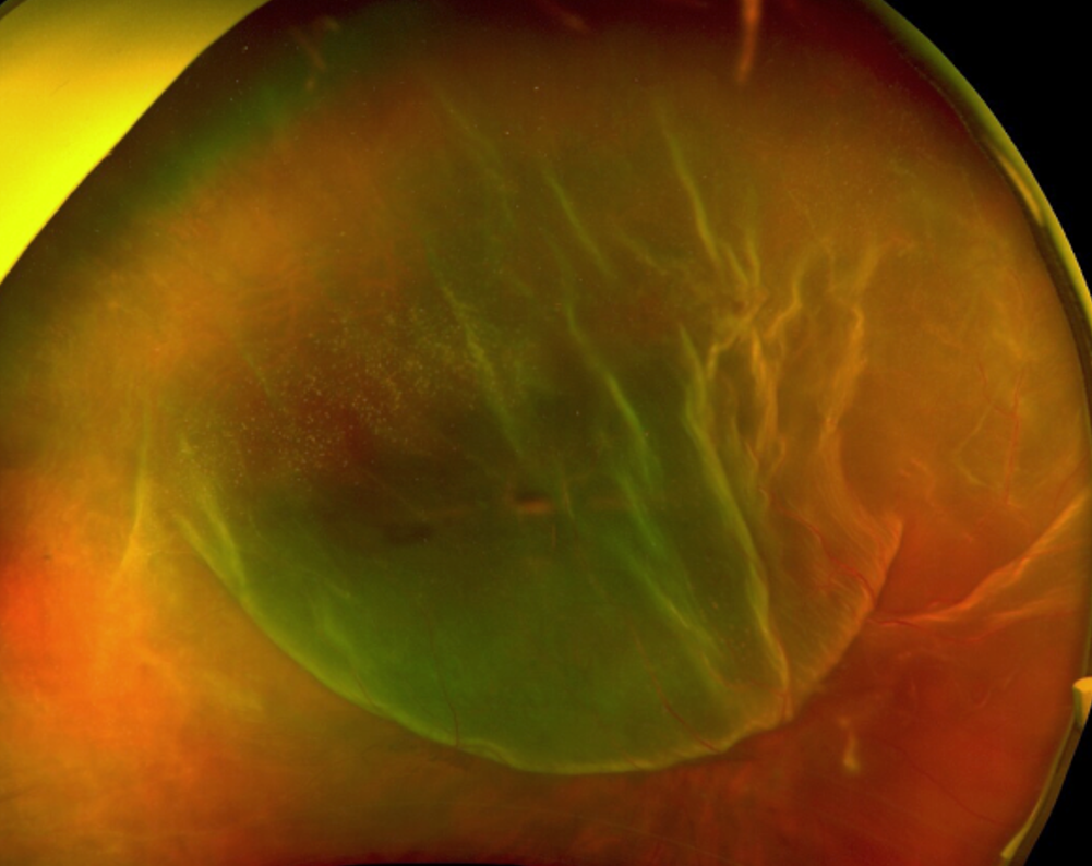
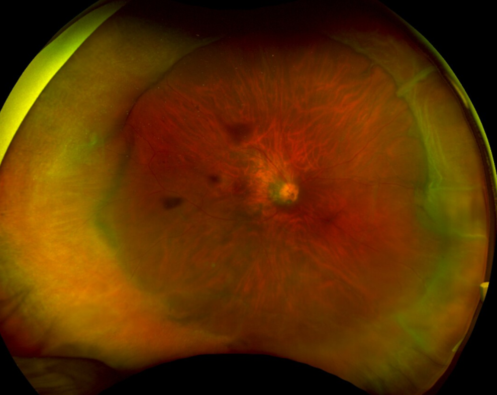
Scleral buckling is mainly used for selected detachments caused by a retinal tear or hole. Your surgeon considers the location and size of the breaks, the condition of the vitreous gel, whether your natural lens is still present, and any scar tissue or previous surgery. It is not the usual treatment for every type of retinal detachment.
This is an extraocular procedure, meaning that buckle-only retinal detachment repair does not require the surgeon to enter the eyeball. It therefore avoids risks associated specifically with intraocular surgical steps, such as cataract progression after vitrectomy and raised intraocular pressure from gas tamponade when no gas is used.
Before recommending surgery, your ophthalmologist examines the retina after using drops to widen the pupil. An ultrasound may be needed if the retina cannot be seen clearly; an OCT scan may help assess the central retina. Not every patient needs every test.[8]
Factors your surgeon may consider
- Location of the retinal breaks. This is the main reason to choose a scleral buckle as the primary intervention. The breaks need to be in the peripheral retina so that, after the buckle is sutured in place, they sit on the indent and are supported by it.
- Lens status. A scleral buckle may be considered in phakic eyes, where the natural lens is still in place, to avoid cataract formation or progression associated with vitrectomy.
- Age and vitreous condition. A buckle may be considered in younger patients, particularly when the vitreous gel has not separated from the retina.
- Retinal dialysis. This is a tear where the outer edge of the retina separates from its attachment. Some detachments caused by this type of tear may be treated with scleral buckling.
- Combined surgery. A buckle may be added to vitrectomy when additional external support is considered useful based on the location or extent of the retinal breaks and other features of the detachment.
Research supports considering whether the natural lens is still present. In the 2007 SPR study of selected, moderately complex detachments, patients with their natural lens had greater improvement in vision with buckling, while those with an artificial lens had better initial reattachment results with vitrectomy.[2] The study does not establish which operation is best for every patient today.
Scleral buckle vs vitrectomy
Scleral buckling and vitrectomy can be used separately or together. Neither is the best choice for every retinal detachment.[2][6] The table compares practical features; your examination findings determine which approach is suitable.
| Consideration | Scleral buckle | Vitrectomy |
|---|---|---|
| Surgical approach | Supports the retina from outside the eye | Uses small instruments inside the eye |
| Vitreous gel | Usually remains in place | The vitreous is removed so that the retina can be accessed |
| Retinal break treatment | Usually freezing treatment called cryotherapy; laser can also be performed postoperatively | Laser is performed during vitrectomy to directly seal the edges of the retinal break |
| Lens considerations | May be considered in eyes that still have their natural lens | Can be performed in phakic eyes with a natural lens and pseudophakic eyes after cataract surgery |
| Cataract progression | The external buckle itself does not induce cataract; cataracts can still develop for other reasons or after combined surgery | Can cause early cataract formation and speed up cataract progression in an eye with a natural lens |
| Refractive effect | Can increase short-sightedness and astigmatism | Does not indent the eye wall in the same way as a buckle |
| Head positioning | Usually no special positioning for buckle-only surgery without gas; instructions may differ if gas or combined surgery is used | Head positioning, often face down, may be required in the first few days, depending on the procedure and tamponade |
| Air travel | No gas-related restriction if no bubble is used; no flying while any intraocular gas or air bubble remains | No flying while an intraocular gas or air bubble remains |
| Combined treatment | May be combined with vitrectomy | May be combined with a buckle |
On smaller screens, swipe horizontally to view the full table.
Your surgeon will explain the recommended operation and relevant alternatives. A gas-bubble procedure called pneumatic retinopexy may suit some detachments. Laser or freezing alone can treat retinal tears before a detachment develops, and laser may be suitable for selected small detachments. These options do not replace surgery for every detached retina.[8]
How scleral buckle surgery is performed
-
Anaesthesia and preparation
Surgery may use regional anaesthesia to numb the eye, often with sedation to help you relax and remain calm during the procedure. General anaesthesia can also be chosen.
-
Accessing the sclera
The conjunctiva, the outermost covering of the eyeball, is opened to access the white surface of the eye, called the sclera.
-
Placement of the buckle
A silicone sponge, band or buckle element is placed on the sclera, behind the muscles around the eyeball, and secured to the sclera with sutures. The configuration depends on the retinal breaks: a segmental buckle supports one area, while an encircling band extends around the eye.
-
Drainage when required
Subretinal fluid may be drained through a controlled opening in the sclera.
-
Checking support and treating the break
Using a light source and lens, called binocular indirect ophthalmoscopy, the surgeon locates the retinal breaks and checks that they sit on the indent. Cryopexy is then applied around the retinal break to scar the edges.
-
Closing the conjunctiva
The conjunctiva is usually closed with sutures that will fall out on their own. Antibiotics are injected at the end of surgery before the eye is padded.
Does the buckle stay in place?
In general, the buckle is left in place for life. Removal is considered only when a later problem, such as exposure, infection or persistent discomfort, makes it necessary.
Recovery after scleral buckle surgery
First few days
Redness, mild swelling, grittiness and soreness are common. Vision is usually blurry, but the darkened area caused by the detachment should be brighter.
You will use postoperative antibiotic and anti-inflammatory eye drops. You will also be taught how to care for the eye, mainly to keep it clean and avoid rubbing.
The following weeks
Redness and discomfort gradually reduce. Restrictions typically include swimming, strenuous exercise and heavy lifting.
When you return to work depends on your occupation and how the eye is healing. Follow-up appointments are used to confirm that the retina remains attached.
Vision over the following months
Once the retina is reattached, visual function should gradually improve. However, vision may take weeks or months to stabilise, especially if the macula was detached. The final vision may not return to its previous level.
A successfully reattached retina does not guarantee full visual recovery. The outcome depends particularly on whether the macula, the central area used for reading and fine detail, was detached and for how long.[1]
A buckle may make the eye more short-sighted or cause astigmatism, affecting how clearly it focuses. Your glasses prescription will be reassessed once the eye has recovered, and you will then be able to update your spectacles.
Positioning and flying
If a scleral buckle is performed without gas tamponade, there is no need to adopt face-down positioning and there are no gas-related restrictions on air travel. Your surgeon can advise on travel timing around your recovery and follow-up appointments.
If gas or air has been placed inside your eye
Do not fly or travel to high altitude until the bubble has fully disappeared and your surgeon confirms it is safe.
Tell every doctor, anaesthetist and dentist treating you that you have a bubble in your eye.[7]
Outcomes and the possibility of further surgery
About 90% of acute retinal detachments can be repaired with one operation.[1] This describes reattachment of the retina, not a 90% chance of recovering normal sight. A small proportion of patients may require additional procedures after surgery, such as laser or gas injections.
Understanding the outcome
When reviewing outcomes, it helps to separate three questions:
- Anatomical outcome: whether the retina is reattached.
- Visual outcome: how much useful sight returns.
- Further treatment: some patients require another operation. This may involve repeat surgery with a buckle, vitrectomy, or a combined approach.
The main factors influencing visual recovery are whether the macula had detached before surgery, how long the retina was detached, and the overall condition of the retina.
Risks and possible complications
Possible postoperative effects
- Soreness, redness and swelling around the eye
- Temporary blurred vision
- A change in glasses prescription, such as increased short-sightedness or astigmatism
- Temporary double vision
Possible complications
- Raised eye pressure
- Infection around the buckle and, rarely, inside the eye
- Bleeding around the buckle
- The buckle becoming exposed through its covering tissue, moving out of position or pressing through nearby tissue in the long term
- Persistent discomfort or double vision
- The retina detaching again, sometimes because scar tissue contracts and pulls on it. This would require further operations to restore the retina.
Some complications require medicines, buckle removal or another retinal operation. Serious complications can cause permanent loss of vision. Your surgeon will explain the risks most relevant to your eye.
Cost of scleral buckle surgery in Singapore
The cost of scleral buckle surgery depends on the buckle configuration, the type of anaesthesia, the operating facility, the complexity of the case, and whether vitrectomy or another procedure is performed at the same time.
The Ministry of Health lists a retinal-detachment buckling operation involving a single buckle element under TOSP code SL801R, Table 5C.[3] Other buckle configurations, complex detachments or combined surgery may use a different code. The code must match the operation performed.
MediSave eligibility and insurance payouts are subject to different rules and limits. Ask the clinic for a written estimate and the applicable procedure code, and check your MediShield Life or Integrated Shield Plan benefits with your insurer where relevant. Private treatment may leave additional out-of-pocket costs.
Read more in the retinal surgery cost section .
Our retina team

Dr Lee Shu Yen
Senior Consultant Ophthalmologist and Vitreoretinal Surgeon
MBBS, MMed (Ophthalmology), MRCSEd, FRCSEd (Ophthalmology), FAMS
Dr Lee is a fellowship-trained vitreoretinal surgeon and former Head of the Surgical Retina department at the Singapore National Eye Centre. Her clinical interests relevant to this page include retinal detachment surgery, giant retinal tears, retinal complications of high myopia and combined vitreoretinal procedures.
Frequently asked questions
Does a scleral buckle stay in the eye permanently?
In most cases the buckle is left in place. It is removed only if a later problem, such as exposure, infection, migration or persistent discomfort, makes removal necessary.
Can a scleral buckle be seen from outside the eye?
A buckle is usually covered by the conjunctiva, the thin tissue over the white of the eye, and is not normally noticeable in everyday interaction. A visible buckle, a new lump or persistent irritation should be checked by your ophthalmologist.
Can I feel the buckle after surgery?
Some sensation of pressure or a mild foreign-body feeling can occur in the early recovery period. For most patients this settles with time, although the experience varies between individuals.
Will scleral buckle surgery change my glasses prescription?
Yes. A buckle can change the eye's shape, increasing short-sightedness or causing astigmatism. Your prescription can be reassessed once it has stabilised. The amount of change varies; glasses cannot reverse retinal damage.
Will I need to lie face down after surgery?
Specific head positioning, such as face-down posturing, is not automatically required after every buckle operation. It may be prescribed if a gas bubble or another combined procedure is used. Your surgeon will explain what your recovery involves.
When can I fly after scleral buckle surgery?
If a gas or air bubble was used, do not fly until it has fully disappeared and your surgeon confirms it is safe. Cabin-pressure changes can make the bubble expand and dangerously raise eye pressure. If no bubble was used, ask your surgeon when flying is appropriate for your recovery and follow-up arrangements.
Can scleral buckle and vitrectomy be performed together?
Yes. A combined procedure is used in selected cases where external support and internal access to the retina are both considered useful.
Can the retina detach again after scleral buckle surgery?
Yes. The retina can detach again, even after an initially successful repair. Keep your follow-up appointments. New flashes, a sudden increase in floaters, a returning shadow or worsening vision need urgent assessment.
Can I still develop a cataract after scleral buckle surgery?
Yes. Cataracts can still develop after buckle surgery. Buckle-only surgery generally has a lower risk of cataract progression than vitrectomy in eyes with a natural lens.[2] Your risk also depends on age, existing lens changes and whether vitrectomy or gas is used with the buckle.
When might a scleral buckle need to be removed?
Removal may be considered if the buckle becomes exposed, infected, migrates, causes persistent pain or is linked to persistent double vision that cannot be corrected in other ways.
Arrange a retinal assessment
A retinal detachment needs prompt specialist assessment because delaying necessary treatment can lead to permanent vision loss. Your surgeon will explain the recommended procedure, its risks, alternatives and the likely consequences of delaying or declining treatment.
The timing of surgery depends on the findings; do not wait for symptoms to worsen.
References
- SingHealth. Retinal Detachment — causes, symptoms and treatment.
- Heimann H, et al. Scleral buckling versus primary vitrectomy in rhegmatogenous retinal detachment: a prospective randomized multicenter clinical study. Ophthalmology. 2007;114(12):2142–2154.
- Ministry of Health, Singapore. SL801R — retinal detachment resection or buckling operation or revision operation with single plomb. TOSP Table 5C.
- American Academy of Ophthalmology EyeWiki. Scleral Buckling for Rhegmatogenous Retinal Detachment.
- Advanced Eye Clinic & Surgery. Retinal surgery page — published summary of Dr Lee Shu Yen's APVRS 2026 presentation on retinal detachment in high myopia and posterior staphyloma.
- Znaor L, Medic A, Binder S, et al. Pars plana vitrectomy versus scleral buckling for repairing simple rhegmatogenous retinal detachments. Cochrane Database of Systematic Reviews. 2019;3:CD009562.
- Royal College of Ophthalmologists. Ophthalmic Safety Alert — Do not use nitrous oxide when there is gas in an operated eye. 18 December 2018.
- American Society of Retina Specialists. Retinal Detachment — patient information.